A fetal echocardiogram is an imaging test technique that allows the doctor to evaluate the structure and function of a developing baby in the womb. This is an essential method of monitoring the condition and cardiovascular health of high-risk babies. From there, the doctor can advise and find the right treatment if detecting an abnormality.
Monitoring the health and development of the fetus is extremely important during pregnancy. Conventional fetal ultrasound can help an obstetrician make an overall assessment of the overall development of a fetus. However, if the pregnant mother is in the group of risks that can affect the cardiovascular health of the fetus, an echocardiogram of the fetus is necessary.
So what is this form of ultrasound in pregnancy? What are the benefits and risks of this method for pregnant women and babies? Will test results be enough to help diagnose and provide future treatment direction? Join aFamilyToday Health to find out information about this imaging method through the article below!
What is echocardiography?
This fetal echocardiogram is a method of using sound waves to create an image of your baby's heart. This imaging test is similar to a conventional ultrasound, is painless and allows the doctor to see the structure and ability of the heart to function.
If necessary, doctors usually prescribe this method during the second trimester, between weeks 18 and 24. The frequency of accurate diagnosis of severe congenital heart disease in the fetus is more than 90%.
Why do you need an ultrasound?
According to statistics from the Ministry of Health , each year our country has about 8,000-10,000 children with congenital heart disease at birth. This proportion accounts for 0.8% of the number of babies born a year. Of which, 50% of children with severe congenital heart disease, but only half have surgery, the rest have to live with the disease and face the risk of death every day.
This ultrasound assists the doctor in assessing the cardiovascular condition of the fetus, such as heart rate, fetal heart function. Doctors also recommend that this method should be included in prenatal diagnosis to help early detect serious heart defects, help timely intervene treatment right from the early stages of pregnancy.
However, not all pregnant mothers need this form of ultrasound. Usually, basic fetal ultrasound also helps the doctor observe the development of all 4 heart chambers in the fetus. Women in the following high-risk groups will need an additional fetal echocardiogram:
Family history of cardiovascular disease
Pregnant mothers have health problems that are at risk for the fetal heart, such as rubella, type 1 diabetes , lupus or phenylketouria
Alcohol, stimulant use during pregnancy
Use or be exposed to certain medications that can cause heart defects, such as epilepsy medications and prescription acne medications
The fetus has a genetic disorder
Abnormalities found in conventional fetal ultrasound
The fetal heart cannot be seen clearly through periodic obstetric ultrasound ...
A fetus in a high-risk group for congenital heart disease will also be prescribed an echocardiogram by the doctor, such as:
Fetal heart arrhythmia
Suspected twins exchange syndrome or multiple pregnancy
Narrowing opacity increased during the first trimester
Edema of the placenta is not hereditary
Abnormalities in the heart
Chromosomal abnormalities: umbilical hernia , nape edema, duodenal atrophy, diaphragmatic hernia ...
The right time to do an echocardiogram of the fetus
During fetal development, the heart begins to form and beat about 22 days after conception, before the pregnant mother even realizes she is pregnant.
Then, by weeks 6–7, the heartbeat begins to appear and modern ultrasound techniques can hear the fetal heartbeat. However, in the early stages, the heart has not yet divided and developed into well-defined chambers and heart valves. From week 20 onwards, the fetal heartbeat becomes stronger, as long as the ear is pressed against the abdomen to be heard. If the fetal heartbeat is louder and easier to hear, the fetus is developing normally and healthy.
If possible, this ultrasound can be performed around 8-10 weeks of pregnancy to detect cardiac abnormalities (if any) early. As usual, an echocardiogram is done around 18-24 weeks.
The process of echocardiography
What should you prepare before having an ultrasound?
In general, you don't need to do any special preparation before having an ultrasound. Pregnant mothers can still eat and drink normally before and do not need to drink as much water to fill the bladder as other ultrasound scans. Note: Do not use cream or powder on the stomach on the day of the scan.
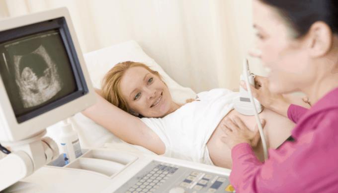
How is an echocardiogram performed?
This test will be done in a dark room and you must stay in the hospital bed. The ultrasound process of the fetus is similar to a regular obstetric ultrasound. Your doctor will apply a gel to your abdomen to help transmit sound waves from the transducer of the fetal echocardiogram to the fetus's heart and respond back. The medical staff will move the probe around the abdomen to get an image of the fetal heart from a variety of angles.
During this type of ultrasound, you may feel a little pressure on your abdomen from the transducer, but the process is really painless.
There are many different ways to help an echocardiogram so that the image is as detailed as possible:
2D ultrasound (two-dimensional / bi-plane ultrasound): shows the structures and functions of the heart at the time of the ultrasound.
Doppler ultrasound: used to measure the rate of blood flow in the chambers and valves of the heart.
An ultrasound of the fetal heart may also be performed through the vagina. At that time, you must undress from the waist down and lie on the examination table. A medical staff will insert a small vaginal probe into the body. This probe also uses sound waves to create an image of the fetal heart. Vaginal echocardiography is usually done early in pregnancy. The doctor can see a clearer picture of the fetal heart through this method.
To get a complete picture of the fetal heart, an ultrasound can take anywhere from 30 minutes to 2 hours. Sometimes, the posture and position of the fetus make it difficult to see the image of the fetal heart. At that time, it will take longer for medical staff to get results.
When will you get your fetal echocardiogram?
Usually, your baby's echocardiogram will be available on the same day you have this imaging test. Sometimes, pregnant mothers need to have an ultrasound again if the results are not clear.
The fetal heart rate of a normal healthy fetus will range from 120 to 160 beats / minute. During fetal movements, the heart rate has the potential to increase up to 180 beats / min. However, if the heart rate rises above this number, it could be a warning sign of serious conditions in the fetus.
Can fetal echocardiograms cause complications for mother and baby?
According to records so far, there is no risk of an echocardiogram of the fetus because this method uses only ultrasound technology and no radiation. Hence, this is a non-invasive imaging test that is safe for both mother and baby.
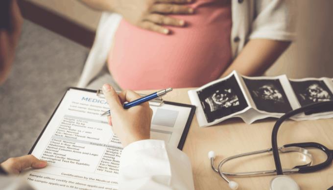
What do fetal echocardiograms mean?
During pregnancy visits, doctors will base on ultrasound results to monitor the normal development of the fetus, especially the cardiovascular system.
If the doctor finds that there are abnormal heart defects, heart rate or other related problems, the pregnant mother will need to do some more tests, such as fetal MRI or high-intensity ultrasound.
Sometimes, you will need more than one fetal echocardiogram or additional tests if the doctor senses that something is wrong with the fetal development.
However, the results of an ultrasound of the fetal heart cannot help detect all the health problems of the baby in the abdomen. Certain conditions, such as having an oval hole in the heart, will be difficult to notice, even with the most advanced, modern equipment.
The doctor can only carefully observe the test results and make the most suitable diagnosis. If there is an abnormality, doctors will rely on it to plan treatment for the baby after birth, such as orthopedic surgery to restore function to the heart. Also thanks to this method, you will receive advice and support from your doctor, helping you to make the best and most appropriate decisions.