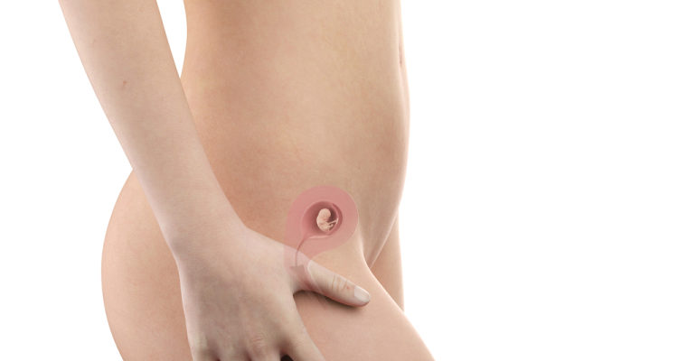
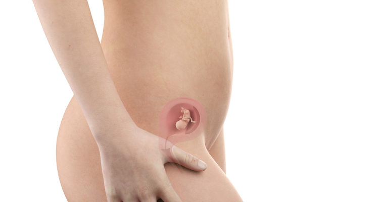
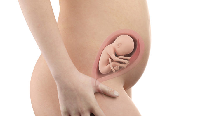
During pregnancy, pregnant mothers will be assigned to do routine tests to ensure the health of you and your baby.
Among the routine tests are those you only need to do once, some that need to be done regularly every time you go to prenatal visit. In addition, based on the gestational age, health status, medical history, the amount of the fetus you are carrying, the results of previous pregnancy tests ... that the doctor may appoint a pregnant mother as a number of additional tests such as amniocentesis , sampling of the chorion ...
Routine pregnancy exams and tests
During pregnancy, pregnant mothers are usually prescribed to perform the following examinations and tests:
1. Check the weight
Not only reflects the health of the pregnant woman but the weight of the pregnant mother also shows the development of the fetus in the womb. Therefore, pregnant mothers are often asked to check their weight every time they go to prenatal care.
Weight gain of pregnant mothers is estimated based on weight before pregnancy.
If you have a normal weight before pregnancy, i.e. body mass index (BMI) is between 18.5 and 24.9, the ideal weight gain for a mother is 10-12 kg, as follows:
First 3 months: Increase 1 kg
Middle 3 months: Increasing 4-5 kg
Last 3 months: Increase 5 - 6 kg
If pregnant women are underweight (BMI: <18.5), during pregnancy the weight gain should reach about 1/4 of the weight before pregnancy, usually 13-18 kg.
If before pregnancy, the mother is overweight, obese (BMI 25 and above): ideal weight gain is 15% of pre-pregnancy weight, usually 7-11 kg.
In case of twins, pregnant mothers need to increase about 16 - 20.5 kg.
2. Check your blood pressure

Every time you go to prenatal check-up, pregnant mother will be checked blood pressure. This is a painless, non-invasive test. Through these measurements, your doctor will determine if you have high blood pressure.
High blood pressure is one of the major risk factors for preeclampsia . In addition, high blood pressure can seriously affect the kidneys and liver of pregnant women. The early detection of high blood pressure during pregnancy helps you to be properly treated, preventing bad complications.
3. Examine the pelvic area
Checking the pelvic area of pregnant mothers helps doctors partly assess the size and position of the vagina, cervix, uterus and ovaries.
The doctor or nurse will insert one or two gloved fingers of one hand into your vagina, with the other hand pressed against your lower abdomen. Your doctor will gently press down on your abdomen and move your fingers around inside the vagina so you can feel the size, shape and texture of the uterus and ovaries. During this procedure, doctors can detect any abnormal growths.
4. Urine test
This is also the same test that you have to do every time you go for prenatal care. This test helps doctors assess if you have a bladder, kidney infection or excess protein in your urine ( proteinuria ). Having too much protein in the urine is a warning sign of preeclampsia that pregnant women need to pay attention to.
5. General blood test for the first quarter
The technician will take your blood from a vein in your arm to be tested. You may feel a bit of pain.
This test will check the liver function of the pregnant mother, check if you are infected with viruses, sexually transmitted diseases, hepatitis B, HIV or not. In addition, through test indicators, doctors will evaluate whether pregnant women are at risk of diabetes, anemia or not, blood type and Rh factor of pregnant mothers (a measuring condition. compatibility between the mother's blood and the baby's blood).
If the test results show that the pregnant mother is in a state of anemia, the doctor can advise on how to eat or give medicine to improve the situation.
The determination of blood type is very important, in case the pregnant mother has an emergency, she will have blood type for timely transfusion.
6. Screening for risk of birth abnormalities

Pregnant mothers are usually indicated to do the following tests:
Combined test: done in the first quarter (11-13 weeks 6 days), screening for Down syndrome, Edward, Patau syndrome, X chromosome abnormality
Triple test: done in the second quarter (15 - 20 weeks), screening for Down syndrome, Edward syndrome, neural tube defects
NIPT: performed from 10 weeks of pregnancy, screening for the risk of the above syndrome, with accuracy of 99% for Down disease.
7. Group B streptococcal infection screening test
This test is done to look for a bacteria (GBS) that causes pneumonia and other serious infections in a newborn. Pregnant mothers will conduct this test in the period from 35 to 37 weeks of pregnancy. To do this test, the doctor swab the fluid in your rectum and vagina with a cotton swab and put it in a test tube for the technician to check. The execution will not cause pain.
If the test result is positive for bacteria, the doctor will give the pregnant mother antibiotics during labor to help prevent infection for the baby.
8. Measure the height of the uterus
From the 16th week of pregnancy, each time you go for prenatal check-up, your doctor will take a uterine height measurement to evaluate the development of the fetus. Usually, the measured length (in cemtimet) corresponds to gestational age (in weeks).
9. Test for sugar tolerance
If you are in the high-risk group for diabetes, your doctor will order you to have this test very early in the early stages of pregnancy.
If you're at no risk, between 24 and 28 weeks of pregnancy, you are usually ordered to have this test to check your blood glucose levels to screen for gestational diabetes.
Pregnant mothers are asked to fast for 8-10 hours before the test (only drink water)
First blood draw when hungry
After the first blood draw, the mother elected to drink 75g glucose. 1 hour after drinking sugar, pregnant mother will receive blood for the second time
The 3rd blood draw is 2 hours after oral sugar, ie 1 hour after the 2nd blood draw.
Results are said to be normal when all 3 readings are normal.
10. Ultrasound
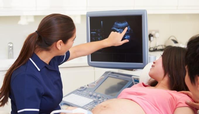
During an abdominal ultrasound, the ultrasound doctor will apply a gel layer to the pregnant womb and move the probe of the ultrasound device on the pregnant womb to get pictures of the fetus, amniotic fluid, adhesion position. of the placenta ... In the case of an ultrasound of the vaginal probe, the doctor will put a probe of about more than 1 finger gently into the vagina to examine the cervix, blood vessels or closely look at the fetal heart during pregnancy. head.
Often pregnant mothers will be assigned an ultrasound right the first time they go to prenatal check-up to check the grip position, size of the pregnancy bag ...
If there is a high risk (multiples of pregnancy, pregnancy when old, each cesarean section, have health problems ...), pregnant mothers may be prescribed ultrasound multiple times.
This is a non-invasive test that uses sound waves to create pictures of the fetus, so it is not painful. Important ultrasounds that a pregnant mother must perform during pregnancy include:
♥ Week 12-14 of pregnancy
Ultrasound checks the number of fetuses, the number of placenta, the amniotic fluid ...
Ultrasound checks the nape of the nape of the neck, which helps to screen for Down syndrome, some defects. Determine the gestational age based on the length of the buttock, give the due date
Uterine Doppler ultrasound helps screen for risk of pre-eclampsia
♥ Weeks of pregnancy 18 - 26
Ultrasound checks for structural and morphological abnormalities of the fetus
Detect abnormalities such as cleft palate, cleft palate, defects in spine, missing or excess fingers ...
Observing the placenta, amniotic fluid, umbilical cord ... to assess the risk of preterm birth for timely intervention
Measure the biological indicators of the fetus to evaluate the development of the baby.
Arterial Doppler ultrasound helps to screen for the risk of pre-eclampsia, evaluate fetal growth
Ultrasound to measure the hole in the cervix: screening for open-waist uterine pathology.
♥ The 32nd week of pregnancy - end of pregnancy
Measure the biological indicators of the fetus to assess the growth of the baby, to see if the baby is at risk of underweight, malnutrition or not
Check the fetal circulation through Doppler ultrasound of the umbilical arteries, the middle cerebral artery, the venous duct ... to assess the risk of the baby having nutrient deficiencies or not.
Check for abnormalities in the development of organs and organs in the fetus such as flat brain, intestinal obstruction, duodenal stenosis ...
Uterine Doppler ultrasound helps screen for risk of pre-eclampsia.
11. Pre-eclampsia risk screening kit
This test suite includes 2-arm average arterial blood pressure measurement, uterine artery Doppler ultrasound, blood test measuring SfLT1 / PlGF. The results help to predict the mother's risk of developing this pathology during pregnancy and thus taking preventive medicine throughout pregnancy.
Implementation time: It can be done at 1 of 3 times in the first quarter, second quarter or third quarter. If the doctor assesses the pregnant mother is at risk of this disease, you should do it right in the first quarter for early prevention.
12. Non-stress test
The Non-stress test measures the baby's heart rate and checks if the baby is getting enough oxygen. Pregnant mothers are usually prescribed to perform tests in the last trimester of pregnancy.
To conduct the test, the pregnant mother will lie comfortably in bed, the nurse will attach the heart rate monitor to your pregnant belly. In addition, the pregnant mother will be given a device and asked to press each time she feels the baby kick or move.
Note that before performing this test, pregnant mothers need to eat well, do not have excessive physical activity ... to not affect the results. During the test, pregnant women should not sleep.
13. Counting fetal movements
This is a test that you can do on your own at any time and place, involving counting the number of fetal movements you feel.
You should start with a pregnancy motion count from around 20 weeks of pregnancy and not be hungry. Maintain a motion count 3 times per week. Usually babies pedal 3-4 times in 1 hour, but sometimes the baby has sleep phase. If your baby does not kick enough times in 2 hours, get up and go back and forth and drink a glass of water or eat something, then count again for 1 more hour. If your baby still kicks a little, go to the hospital immediately to check. Because this can be a warning sign that your baby is in danger.
In addition, if the screening tests suggest that the fetus is at high risk of birth defects, the mother may need to do more invasive tests to diagnose conditions such as: placenta biopsy, puncture amniotic fluid detection.
Useful when pregnant mothers perform routine screening and testing
During routine tests you will learn the following useful information:
High blood pressure: A marker of pre-eclampsia risk. A very dangerous situation.
Pregnant mothers have gestational diabetes or not in order to promptly intervene to avoid complications affecting the health of you and your baby. Gestational diabetes usually goes away after birth but can increase your risk of type 2 diabetes.
Birth defects and genetic abnormalities: Many birth defects and genetic abnormalities are not caused by pregnancy. However, they can be detected during pregnancy, including Down syndrome, cystic fibrosis and spina bifida.
Striker placenta : This happens when the placenta covers the mother's cervix. This is a condition that can cause serious bleeding during pregnancy and childbirth.
The fetus is not in the dominant position : Usually at the end of pregnancy, the fetus's head is usually in the down position of the pregnant mother's pelvis. If the fetus is in the buttock position (buttocks bent over the pelvis instead of the head) or transverse (the baby is horizontal in the uterus), this interferes with the normal delivery of the pregnant mother, increasing the risk of having cesarean section.
Pregnancy is a sensitive stage, so to ensure the health of the pregnant mother and the development of the fetus, pregnant mothers should fully perform antenatal care and do tests as prescribed by a doctor. This helps to promptly intervene if a bad situation occurs.
You may be interested in the topic:
Falls during pregnancy and measures to ensure the safety of pregnant mothers
Premenstrual psychosis: Severe and few people know
42 weeks of pregnancy not yet born: Pregnant mothers do not worry too much


